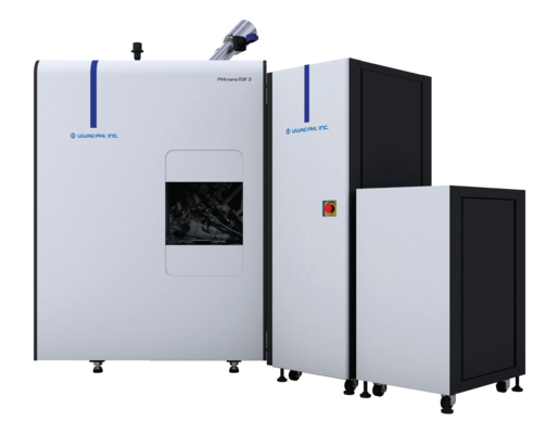
PHI’s patented Parallel Imaging MS/MS mass spectrometer provides superior sensitivity, low spectral background, unique ability to image highly topographic surfaces, high mass accuracy and mass resolution, and unambiguous peak identification with parallel tandem MS imaging capability. The PHI nanoTOF 3 can be configured with a wide variety of options to optimize performance for organic materials, inorganic materials, or both, depending on customer requirements.
NEW Pulsed Dual-Beam Charge Neutralization For Truly Turn-Key Insulator Analysis
- Self-regulating charge neutralization by low energy electrons and low energy inert gas ions
- Artifact-free chemical imaging by neutralization of the position-dependent surface charges
NEW Bi Cluster Emitter With Smaller Beam Diameter For Improved High-Throughput HR2 Imaging
- The most utilitarian analysis mode with < 500 nm chemical characterization
- The HR2 imaging is attained at high analysis beam current so the analysis time is short
- Delayed extraction (DE) is not necessary, so the artifacts of DE are completely avoided
Confident Chemical & Molecular Analysis Of Curved, Rough & Charging Samples
- Superior angular acceptance and depth-of-field of the mass spectrometer
- Artifact-free chemical imaging of ex situ and in situ FIB-sectioned samples
- Superior imaging of materials by correcting the position-dependent surface potential
- Delayed extraction (DE) is not necessary, so the artifacts of DE are completely avoided
NEW Automated Stage & In-Vacuum Parking for High-Throughput Sample Handling & Unattended Analysis
- Equipped with an automatic sample transfer mechanism that has been proven in more than 300 Q-series XPS instruments
- Sample sizes up to 100 mm x 100 mm, and the analysis chamber has a built-in parking mechanism as standard
- Combined with the Queue Editor, the system allows continuous automated measurements of large numbers of samples
Tandem Mass Spectrometers For Rapid Imaging And Accurate Peak Identification
- No data is ever discarded; integrated and uncomplicated TOF-SIMS (MS1) and tandem MS (MS2) imaging
- Ultra-high transmission for optimized molecular sensitivity and monolayer analysis
- High speed (> 8 kHz) TOF-SIMS (MS1) and tandem MS (MS2) imaging
- High resolution (< 50 nm) TOF-SIMS (MS1) and tandem MS (MS2) imaging
- High energy (> 1.5 keV) collision-induced dissociation (keV-CID) for identification
Solutions For In Situ Characterization Of Advanced Materials
- All operation modes available for automated and unattended analysis
- Cryo- and high-temperature analysis via the Hot/Cold and High-Temperature module options; full 5-axis sample motion is maintained during temperature-controlled analysis
- Advanced 3D imaging with use of optional Ar, O2, Cs, C60, Ar cluster, and Ga-FIB ion beams
- Solid-state electrochemistry (biasing, polarization) experiments with the Static Voltage & Power Cycling option
- Inert Sample Transfer Vessel and Transfer Vessel Adapter (25 mm AES/XPS puck) options
Superior Parallel Imaging MS/MS Analyzer Performance
Integrated tandem mass spectrometers, i.e., Parallel Imaging MS/MS, for lossless collection of TOF-SIMS (MS1) and tandem MS (MS2) data, and with keV-CID for unambiguous peak identification and structural analysis.
Superior depth-of-field and uniform imaging sensitivity on curved or rough surfaces due to the large angular acceptance and band-pass kinetic energy window of the spectrometer.
Low spectral background and highest abundance sensitivity due to the automatic filtering of metastable post-source decay (PSD) ions.
High mass sensitivity for geological, organic and bio-medical applications.
Truly turn-key insulator analysis enabled by the newly patented pulsed dual-beam charge compensation technology.

Parallel Imaging MS/MS
TOF-SIMS is a fast MS imaging tool with consequently low mass accuracy and mass resolution that is an impediment to unambiguous peak identification. PHI’s revolutionary and patented Parallel Imaging MS/MS spectrometer technology obliterates this limitation with an integrated and lossless time-of-flight (TOF) tandem MS (MS2) capability that makes use of high-energy (1.5 keV) collision-induced dissociation (keV-CID). With the tandem MS enabled, a selected precursor ion (monoisotopic selection window) is deflected into the CID cell while the remaining ions continue to the TOF-SIMS (MS1) detector as usual. The precursor ions collide with argon gas in the CID cell causing fragmentation, and the resulting product ions travel to the tandem MS (MS2) detector giving rise to the MS/MS (MS2) product ion spectrum. Similar to the MS1 data, a full MS2 spectrum is collected for each image pixel. Thus, TOF-SIMS and tandem MS data are collected simultaneously, in parallel, and without discarding any ions, from the same analytical volume. All typical TOF-SIMS attributes remain, including high-speed imaging, high sensitivity, and high spatial resolution.
The MS2 product ion spectrum can be used to identify the precursor ion composition directly from the product ion spectrum or by comparison to mass spectral databases. The Parallel Imaging MS/MS spectrometer also improves detection sensitivity for species in which the peak of interest has a mass interference by detection of a unique product ions.

Heat treated PET sample. Thermal-scaled images of MS1 and MS2 ion peaks demonstrating the collection of two spectral data sets from every image pixel and confirming that the PET trimer is localized only to the thermally precipitated surface crystals. Positive ion polarity, 40 μm field-of-view, 10 μm scale bar.

Heat treated PET sample. Composition assignments for the MS2 spectrum obtained by keV- CID fragmentation of the +m/z 577 precursor ion confirm that the observed crystals are composed of ethylene terephthalate trimer.

Automotive polymer sample. MS/MS image (MS2) of the +m/z 481 precursor ion showing a non-uniform distribution of the species on the sample surface.

MS/MS spectrum (MS2) obtained from CID fragmentation of the +m/z 481 precursor ion. Identification of the fragment ions indicates that the precursor ion [M+H]+ is Tinuvin 770, a polymer additive.

Further confirmation of the identity of the precursor ion is made with a positive match to Tinuvin 770 in a commercially available MS/MS database which is included with tandem MS/MS. The spectrum in red is the MS2 spectrum and the blue spectrum is the acquired reference database spectrum of Tinuvin.
HR²
High spatial resolution with high mass resolution
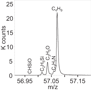
HR² imaging is demonstrated below in the images of micro-organic droplets. In a single analysis, peaks for multiple molecular fragments were observed in the spectrum at m/z 57 with high mass resolution and identified by measurement of their exact mass. The distribution of each species was visualized with a spatial resolution of less than 400 nm. The single measurement that contains all this information was acquired in only six minutes due to the high ion beam current available in the PHI nanoTOF’s HR² imaging mode.


Cluster Source Ion Guns
Multiple cluster source ion gun options
A 20 kV Ar2,500+ gas cluster ion beam (GCIB) option is available for molecular depth profiling organic films. Shown in the figure (right) is a depth profile of a multi-layer polystyrene and poly(2-vinylpyridine) block copolymer film obtained by sputtering with a 5 kV Ar2,500+ gas cluster ion beam.

Depth profiles of a 6 μm-thick aluminosilicate sol-gel on copper by Ar and C60 sputtering. The C60 ion beam does not produce the preferential sputtering artifacts produced by the Ar ion beam.
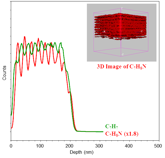
FIB-TOF 3D Chemical Imaging

- TOF-SIMS chemical imaging of in situ FIB-sectioned specimens. This can be performed using the liquid metal ion beam (LMIG) of the base system; however, 3D tomography requires the dedicated Ga-FIB option.
- High mass resolution spectra are collected at every image pixel with uniform sensitivity is enabled by the Parallel Imaging MS/MS spectrometer. Delayed extraction is not used, so the artifacts of delayed extraction are completely avoided.
- User-friendly 3D imaging software with multi-element overlay capability is provided.
3D FIB-TOF images of a solid oxide fuel cell with a 50 x 50 x 10 µm analysis volume show the ability to observe compositional information and physical structures such as voids.
TOF-DR Software for TOF-SIMS Tandem MS Imaging Data Processing
TOF-DR 3.0 is the latest release of PHI’s software for the treatment, processing, presentation and reporting of TOF-SIMS tandem MS imaging data.
The new release includes new or improved capabilities for instant viewing of raw data, peak identification, graphical data presentation and report generation.
Features include:
- Windows 10 64-bit compatibility
- TOF-SIMS MS/MS spectra and 2D/3D image analysis
- Spectral dead time correction
- 2D/3D region-of-interest (ROI) analysis
- Software-guided batch data processing with output to Excel® for plotting and graphical viewing of the raw and normalized data
- Data Review Utility tool for instant and simultaneous observation of peaks, images and profiles with identification tools
- Report Generation tool for easy layout and presentation of data with automated creation of PowerPoint® slides
- PLS_Toolbox® compatibility for full uni- and multi-variate data analysis
- NIST MS/MS® compatibility for tandem MS precursor identification


Brochures
Application Notes
Papers
What Our Customers Say
“The PHI Genesis X-ray Photoelectron Spectroscopy (XPS) instrument has revolutionized our lab's capabilities. Its advanced features and flexibility enable detailed surface material analysis, while PHI's exceptional support ensures smooth operations. The seamless installation and insightful training provided by PHI's knowledgeable field engineers have made this system an invaluable addition to our facilities.”
Quantum Valley Ideas Lab
“PHI's service team is exceptional. Their engineers are professional, reliable, and knowledgeable, always seeking help when needed to ensure reliability. The scientists are equally impressive, providing clear guidance for analysis. PHI's customer service is amazing.”
NY CREATES
“As a global supplier in the automotive, aerospace, and industrial markets, we rely on PHI Auger instruments for customer support, production, and R&D. The PHI 710 Auger, with its state-of-the-art capabilities and excellent customer support, continues to drive our product and process development. The expertise of PHI's service engineers and scientists, along with the flexibility to add specific capabilities, strengthens our long-standing relationship with PHI.”
Manufacturing Customer
“PHI's prompt and willing responses to our VersaProbe system and data analysis concerns have been invaluable. Their excellent application support ensures we always have the help we need.”
University of Virginia
“PHI's TOF-SIMS studies were instrumental in resolving a major issue in our products. Their expertise helped us identify the compromised film, enabling us to successfully address the problem. PHI's support was a significant contributor to our success.”
Semiconductor Contract Customer
Previous Next“We are very pleased with the timely assistance our users have received from PHI's application department over the past six years. The experienced application scientists provide invaluable tips and share application materials promptly, often on the same day. PHI's support has been crucial in expanding our user base and exploring new applications on the system.”
Seagate
Learn how our instruments can help with your applications